Wednesday, March 30, 2011

The Neurobiology of Conscious Intent

Perhaps the seminal component of any clinician’s behavioral repertoire is the ability to understand the conscious motivations and intentions of their clients. This article addresses the work of conscious motivations at the neuroanatomical level.I seldom address the notion of consciousness—let alone motivations—in this column for a very good reason. Nobody really knows what they are or even if there is a “they.” The literature is confusing, but it hasn’t stopped researchers from speculating on possible neuroanatomical and biochemical substrates that undergird the phenomena. Without a broad consensus about what is being studied, there can be no neurons, let alone molecules, for active experimental consideration. After all these years, researchers have yet to isolate an area of the brain solely devoted to the experience of consciousness. There may be none.
Given the importance of these issues to the mental health professions, I revisit the concept of motivations from time to time—but only when the data are conservatively presented, with sober, modest conclusions. The findings described here originate from experiments that have attempted to determine how we voluntarily choose to perform a motor task (action planning). This work requires reviewing background information on association cortices and the neural substrates behind a decision to initiate voluntary action.
Association cortices
Functionally, the cortical regions of the brain and their myriad interlocking circuits can be divided into 3 modules. These consist of front-, back-, and middle-end domains.
• Front-end functional domains are sensory information processing centers. The brain receives input from the eyes, ears, and other sensory systems. It sends the input off to various places for further processing.
• Back-end functional domains involve motor control systems. These systems essentially respond to whatever command the sensory cortices give to it (eg, execute a decision to move).
• The middle-end suite involves nearly everything other than front-end and back-end functional domains. These association cortices generally entail higher processing features and are some of the least understood and the most mysterious parts of the brain.
One such cortex, located in the inferior posterior parietal cortex, is a sensorimotor association region that links sensory stimuli to motor movement. It may even be involved in sensory prediction, which calculates the consequences of a given action through the simultaneous evaluation of input from both sensory (front-end) and motor (back-end) functional domains.
Volitional motor movement
Many of the actions humans initiate on a day-to-day basis seem to depend on a kind of internal free will. This sequence of events (also known as volitional motor movement) gives humans a sense of control: we act because we want to act. That is why researchers use volitional motor movements in their research designs. Researchers interested in volitional behavior study neural prime movers behind decision making.
Exactly what does it mean to want to do something? We do not really know. The events that initiate movement occur in a fairly straightforward sequence (although it depends on the source of the signal). For example, a central processing area with directives for voluntary motor movements pass through a final staging area before the execution of an action. This region is the primary motor cortex.
Research on laboratory animals demonstrates that this cortex decides on a course of action that depends on the source of signals it receives before the execution of that action. One source originates in the premotor cortex. Signals in this area initiate movements in response to a specific external trigger, such as a visual cue.
The second source arises in the presupplementary motor area, which is stimulated when laboratory animals make the same movements mentioned above, but they do not originate from responses to an external source. The movement instead arises spontaneously; a thought is internally generated through intentional actions. There is an observed rapid rise in electrical signals that build up just before the brain executes these actions. This has led to the notion that the presupplementary motor area harbors some kind of readiness potential, a useful function in generating movement (Figure).
In terms of human behavior, complex human brains have many more research issues to solve than standard laboratory animal research can address. One potential confounder is conceptual. With research of this type, scientists often tell subjects to choose (or not to choose) from a variety of options. Is that voluntary? Hardly. This is like saying, “Okay, it’s time to have some voluntary volitional behavior now,” or like runners at a race who respond to the starting gun. Do volitional actions disappear in these experiments with human subjects? Are these subjects simply reacting to commands to respond, not to respond, or to respond however they want? To test volition, researchers should not control the input. Nevertheless the experimenter must, almost by definition.
Wilder Penfield revisited
Another complexity involves engineering. How does conscious intent to move an arm relate to the actual movement of the arm? This could be partially resolved with electrical stimulation mapping in which surgeons create a map of the brain on conscious patients to understand what tissues need to be avoided during certain manipulations (such as resection). No pain neurons exist in the brain. The patient, immobilized in a stereotactic frame, can be consciously interrogated while the surgery takes place. The surgeon applies a gentle electrical current to the open tissue, talks to the patient about what he or she is experiencing, and makes a map that discerns what areas to avoid during cutting. Working primarily with epileptic patients, the legendary Canadian physician Wilder Penfield first performed these techniques.1
This technique has proved to be of great value in understanding volitional components of motor movement. It was discovered almost 2 decades ago that if one stimulates a specific area of the human presupplemental area, the patient will experience a conscious urge to move.2 This gets around the runner’s starting gun problem mentioned previously. An external electrical stimulator supplies a specific quantity of electricity—and a desire to do something is suddenly generated!
As important and well-characterized as these data are, they hardly explain what causes the presupplemental area to generate the signal in subjects not undergoing surgery. Some research findings answer this question and have led to some intriguing results.3,4
When the inferior posterior parietal cortex was stimulated, the patient experienced an urge to move specific body parts. Stimulating one area caused patients to want to move their arms. Another region, the lips. Another region, the chest. This is similar to what one observes in frontal lobes, except that you are nowhere near the frontal lobes. Recall that this is the associative cortex region (a sensorimotor associative area at that), quite distinct from anything observed in the well-characterized general motor areas of the frontal lobes. Was this simply a remote stimulation?
This result showed that the answer would be no. The parietal cortex urges were qualitatively different from those obtained by stimulating parts of the presupplementary cortex. It is well known that if the presupplementary cortex is stimulated at a low current, the urge to act is acquired. However, if the same region is stimulated at high current, actual movement occurs. That’s not what happened in the parietal cortex. The urge was stimulated at low intensities, but movement was never generated at higher ones. Instead, subjects felt that they had already performed some movement.
This is important. The desire to move did not result from subtle motor contractions that may have been generated by motor regions (an alternative idea that has been put forth as a rational explanation for the results in previous experiments). Parietal stimulation never produces muscle activity, regardless of the intensity. The stimulation of the premotor cortex itself produces large-limb movements in subjects, but never the desire to move the limbs. They usually remain unaware that movement has occurred when these regions are stimulated.
These results suggest the presence of 2 specific aspects of conscious intention (however one defines it). One might be the conscious correlation of preparatory motor commands in the presupplemental cortex region, as is clearly observed in laboratory studies of animals. The other might involve sensory prediction of the consequences of those commands, under the domain of the association cortex region. A portion of conscious intent seems to be a specific class of experiences housed within the parietal lobe.
Conclusions
It appears that the parietal lobe contributes to the conscious experience of intention, at least in regard to motor movement. These results cement 1 more brick onto the great construction project that seeks to define intention. But they hardly hint at the overall building.
Pushing the edge of our understanding into the murky world of association cortex only means that future experiments will be trickier to interpret. Electrical stimulation mapping, as good as it is, is necessarily a blunt instrument that stimulates thousands of neurons simultaneously. Not isolated modules, these regions connect to each other in complex, little-understood ways. That the regions produce different behaviors is an important finding but not a defining one.
How do the frontal and motor aspects of volitional experience differ from the parietal, sensory versions? What factors stimulate the parietal lobes in the first place? What about remote effects?
Questions such as these remain to be answered and are just a few of the many that researchers will face as they attempt to define intentional and conscious experiences.
This article originally appeared in the Psychiatric Times.

References
1. Penfield W, Erickson TC. Epilepsy and cerebral localization: a study of the mechanism, treatment and prevention of epileptic seizures (Review). South Med J. 1942;35:222.
2. Fried I, Katz A, McCarthy G, et al. Functional organization of human supplementary motor cortex studied by electrical stimulation. J Neurosci. 1991;11:3656-3666.
3. Haggard P. Human volition: towards a neuroscience of will. Nat Rev Neurosci. 2008;9:934-946.
4. Custers R, Aarts H. The unconscious will: how the pursuit of goals operates outside of conscious awareness. Science. 2010;329:47-50.

The Cell-Cell Structure

 E. coli Bacteria

Life is both wonderful and majestic. Yet for all of its majesty, all organisms are composed of the fundamental unit of life, the cell. The cell is the simplest unit of matter that is alive. From the unicellular bacteria to multicellular animals, the cell is one of the basic organizational principles of biology. Let's look at some of the components of this basic organizer of living organisms.

Eukaryotic Cells and Prokaryotic Cells

There are two primary types of cells: eukaryotic cells and prokaryotic cells. Eukaryotic cells are called so because they have a true nucleus. The nucleus, which houses DNA, is contained within a membrane and separated from other cellular structures. Prokaryotic cells however have no true nucleus. DNA in a prokaryotic cell is not separated from the rest of the cell but coiled up in a region called the nucleoid.

As organized in the Three Domain System, prokaryotes include archaeans and bacteria. Eukaryotes include animals, plants, fungi and protists. Typically, eukaryoitc cells are more complex and much larger than prokaryotic cells. On average, prokaryotic cells are about 10 times smaller in diameter than eukaryotic cells.

Eukaryotes grow and reproduce through a process called mitosis. In organisms that also reproduce sexually, the reproductive cells are produced by a type of cell division called meiosis. Most prokaryotes reproduce through a process called binary fission. During binary fission, the single DNA molecule replicates and the original cell is divided into two identical daughter cells.

Both eukaryotic and prokaryotic organisms get the energy they need to grow and maintain normal cellular function through cellular respiration. Cellular respiration has three main stages: glycolysis, the citric acid cycle, and electron transport. In eukaryotes, most cellular respiration reactions take place within the mitochondria. In prokaryotes, they occur in the cytoplasm and/or within the cell membrane.

The Cell-Cell Structure

There are also many distinctions between eukaryotic and prokaryotic cell structure. The following table compares the cell structures found in a typical prokaryotic cell to those found in a typical animal eukaryotic cell.

Cell Structure Comparison

Eukaryotic and Prokaryotic Cell Structure

Cell Structure Prokaryotic Cell Typical Animal Eukaryotic Cell
Cell Wall Yes No
Centrioles No Yes
Chromosomes One long DNA strand Many
Cilia or Flagella Yes, simple Yes, complex
Endoplasmic Reticulum No Yes (some exceptions)
Golgi Complex No Yes
Lysosomes No Common
Mitochondria No Yes
Nucleus No Yes
Peroxisomes No Common
Cell Membrane Yes Yes
Ribosomes Yes Yes

Animal Cell Structure

Animal cells are typical of the eukaryotic cell, enclosed by a plasma membrane and containing a membrane-bound nucleus and organelles. Unlike the eukaryotic cells of plants and fungi, animal cells do not have a cell wall. This feature was lost in the distant past by the single-celled organisms that gave rise to the kingdom Animalia. Most cells, both animal and plant, range in size between 1 and 100 micrometers and are thus visible only with the aid of a microscope.
Anatomy of the Animal Cell
The lack of a rigid cell wall allowed animals to develop a greater diversity of cell types, tissues, and organs. Specialized cells that formed nerves and muscles—tissues impossible for plants to evolve—gave these organisms mobility. The ability to move about by the use of specialized muscle tissues is a hallmark of the animal world, though a few animals, primarily sponges, do not possess differentiated tissues. Notably, protozoans locomote, but it is only via nonmuscular means, in effect, using cilia, flagella, and pseudopodia.
The animal kingdom is unique among eukaryotic organisms because most animal tissues are bound together in an extracellular matrix by a triple helix of protein known as collagen. Plant and fungal cells are bound together in tissues or aggregations by other molecules, such as pectin. The fact that no other organisms utilize collagen in this manner is one of the indications that all animals arose from a common unicellular ancestor. Bones, shells, spicules, and other hardened structures are formed when the collagen-containing extracellular matrix between animal cells becomes calcified.
Animals are a large and incredibly diverse group of organisms. Making up about three-quarters of the species on Earth, they run the gamut from corals and jellyfish to ants, whales, elephants, and, of course, humans. Being mobile has given animals, which are capable of sensing and responding to their environment, the flexibility to adopt many different modes of feeding, defense, and reproduction. Unlike plants, however, animals are unable to manufacture their own food, and therefore, are always directly or indirectly dependent on plant life.
Most animal cells are diploid, meaning that their chromosomes exist in homologous pairs. Different chromosomal ploidies are also, however, known to occasionally occur. The proliferation of animal cells occurs in a variety of ways. In instances of sexual reproduction, the cellular process of meiosis is first necessary so that haploid daughter cells, or gametes, can be produced. Two haploid cells then fuse to form a diploid zygote, which develops into a new organism as its cells divide and multiply.
The earliest fossil evidence of animals dates from the Vendian Period (650 to 544 million years ago), with coelenterate-type creatures that left traces of their soft bodies in shallow-water sediments. The first mass extinction ended that period, but during the Cambrian Period which followed, an explosion of new forms began the evolutionary radiation that produced most of the major groups, or phyla, known today. Vertebrates (animals with backbones) are not known to have occurred until the early Ordovician Period (505 to 438 million years ago).
Fluorescence Microscopy of Cells in Culture
Cells were discovered in 1665 by British scientist Robert Hooke who first observed them in his crude (by today's standards) seventeenth century optical microscope. In fact, Hooke coined the term "cell", in a biological context, when he described the microscopic structure of cork like a tiny, bare room or monk's cell. Illustrated in Figure 2 are a pair of fibroblast deer skin cells that have been labeled with fluorescent probes and photographed in the microscope to reveal their internal structure. The nuclei are stained with a red probe, while the Golgi apparatus and microfilament actin network are stained green and blue, respectively. The microscope has been a fundamental tool in the field of cell biology and is often used to observe living cells in culture. Use the links below to obtain more detailed information about the various components that are found in animal cells.
  • Centrioles - Centrioles are self-replicating organelles made up of nine bundles of microtubules and are found only in animal cells. They appear to help in organizing cell division, but aren't essential to the process.
  • Cilia and Flagella - For single-celled eukaryotes, cilia and flagella are essential for the locomotion of individual organisms. In multicellular organisms, cilia function to move fluid or materials past an immobile cell as well as moving a cell or group of cells.
  • Endoplasmic Reticulum - The endoplasmic reticulum is a network of sacs that manufactures, processes, and transports chemical compounds for use inside and outside of the cell. It is connected to the double-layered nuclear envelope, providing a pipeline between the nucleus and the cytoplasm.
  • Endosomes and Endocytosis - Endosomes are membrane-bound vesicles, formed via a complex family of processes collectively known as endocytosis, and found in the cytoplasm of virtually every animal cell. The basic mechanism of endocytosis is the reverse of what occurs during exocytosis or cellular secretion. It involves the invagination (folding inward) of a cell's plasma membrane to surround macromolecules or other matter diffusing through the extracellular fluid.
  • Golgi Apparatus - The Golgi apparatus is the distribution and shipping department for the cell's chemical products. It modifies proteins and fats built in the endoplasmic reticulum and prepares them for export to the outside of the cell.
  • Intermediate Filaments - Intermediate filaments are a very broad class of fibrous proteins that play an important role as both structural and functional elements of the cytoskeleton. Ranging in size from 8 to 12 nanometers, intermediate filaments function as tension-bearing elements to help maintain cell shape and rigidity.
  • Lysosomes - The main function of these microbodies is digestion. Lysosomes break down cellular waste products and debris from outside the cell into simple compounds, which are transferred to the cytoplasm as new cell-building materials.
  • Microfilaments - Microfilaments are solid rods made of globular proteins called actin. These filaments are primarily structural in function and are an important component of the cytoskeleton.
  • Microtubules - These straight, hollow cylinders are found throughout the cytoplasm of all eukaryotic cells (prokaryotes don't have them) and carry out a variety of functions, ranging from transport to structural support.
  • Mitochondria - Mitochondria are oblong shaped organelles that are found in the cytoplasm of every eukaryotic cell. In the animal cell, they are the main power generators, converting oxygen and nutrients into energy.
  • Nucleus - The nucleus is a highly specialized organelle that serves as the information processing and administrative center of the cell. This organelle has two major functions: it stores the cell's hereditary material, or DNA, and it coordinates the cell's activities, which include growth, intermediary metabolism, protein synthesis, and reproduction (cell division).
  • Peroxisomes - Microbodies are a diverse group of organelles that are found in the cytoplasm, roughly spherical and bound by a single membrane. There are several types of microbodies but peroxisomes are the most common.
  • Plasma Membrane - All living cells have a plasma membrane that encloses their contents. In prokaryotes, the membrane is the inner layer of protection surrounded by a rigid cell wall. Eukaryotic animal cells have only the membrane to contain and protect their contents. These membranes also regulate the passage of molecules in and out of the cells.
  • Ribosomes - All living cells contain ribosomes, tiny organelles composed of approximately 60 percent RNA and 40 percent protein. In eukaryotes, ribosomes are made of four strands of RNA. In prokaryotes, they consist of three strands of RNA.
In addition the optical and electron microscope, scientists are able to use a number of other techniques to probe the mysteries of the animal cell. Cells can be disassembled by chemical methods and their individual organelles and macromolecules isolated for study. The process of cell fractionation enables the scientist to prepare specific components, the mitochondria for example, in large quantities for investigations of their composition and functions. Using this approach, cell biologists have been able to assign various functions to specific locations within the cell. However, the era of fluorescent proteins has brought microscopy to the forefront of biology by enabling scientists to target living cells with highly localized probes for studies that don't interfere with the delicate balance of life processes.

Cell Division and DNA Replication

In the first lecture, we covered the way science works and especially how the scientific method applies to biology. Then, we looked at the structure of the cell, building a map of the cell - knowing what processes happen where in the cell, e.g., the production of energy-rich ATP molecules in the mitochondria.
In the third part of the lecture, we took a closer look at the way DNA code gets transcribed into RNA in the nucleus, and the RNA code translated into protein structure in the rough endoplasmatic reticulum. Finally, we looked at several different ways that cells communicate with each other and with the environment, thus modifying cell function.
All of that information will be important in this lecture, as we cover the ways cells divide, how cell-division, starting with a fertilized cell, builds an embryo, how genetic code (genotype) influences the observable and measurable traits (phenotype) and, finally, how do these processes affect the genetic composition of the populations of organisms of the same species - the process of evolution.
Mitosis
The only way to build a cell is by dividing an existing cell into two. As the genome (the complete sequence of the DNA) is an essential part of a cell, it is neccessary for the DNA to be duplicated prior to cell division.
In Eukaryotic cells, chromosomes are structures composed mostly of DNA and protein. DNA is a long double-stranded chain-like molecule. Some portions of the DNA are permanently coiled and covered with protective proteins to prevent DNA expression (transcription). Other parts can be unraveled so transcription can occur.
The number of chromosomes is different in different species. Human cells possess 23 pairs of chromosomes. Prior to cell division each chromosome replicates producing two identical sister chromosomes - each eventually landing in one of the daughter cells.
The process of DNA replication - the way all of the DNA code of the mother cell duplicates and one copy goes into each daughter cell - is the most important aspect of cell division. It is wonderfully described in your handout and depicted in the animation. Other cell organelles also divide and split into two daughter cells. Once the process of DNA replication is over, the new portion of the cell membrane gets built transecting the cell and dividing all the genetic material into two cellular compartments, leading the cell to split into two cells.
a1%20mitosismeiosis.jpg 
Meiosis
Meiosis is a special case of cell division. While mitosis results in division of all types of cells in the body, meiosis results in the formation of sex cells - the gametes: eggs and sperm. Mitosis is a one-step process: one cell divides into two. Meiosis is a two-step process: one cell divides into two, then each daughter immediately divides again into two, resulting in four grand-daughter cells.
Each cell in the body has two copies of the entire DNA - one copy received from the mother, the other from the father. Fertilization (fusion of an egg and a sperm) would double the chromosome number in each generation if the egg and sperm cells had the duplicate copy. Meiosis ensures that gametes have only one copy of the genome - a mix of maternal and paternal sequences. Such a cell is called a haploid cell.
Once the egg and a sperm fuse, the resulting zygote (fertilized egg) again contains double dose of the DNA and is called a diploid cell. Thus the resultant zygote inherits genetic material from both its father and its mother. All the cells in the body except for the gametes are diploid. Sexual reproduction produces offspring that are genetically different from either parent.
DNA Replication
DNA replication is a complex process of duplication of the DNA involving many enzymes. It is the first and the most important process in cell division. Please read the handout (BREAKFAST OF CHAMPIONS DOES REPLICATION by David Ng) to appreciate the complexity of the process, but you do not need to memorize any of the enzymes for the exams. Also, it will help your understanding of the process if you watch this animation.

Algorithms in Structural Molecular Biology and Proteomics

Some of the most challenging and influential opportunities for Physical Geometric Algorithms (PGA) arise in developing and applying information technology to understand the molecular machinery of the cell. Our recent work (e.g., [1-20]) shows that many PGA techniques may be fruitfully applied to the challenges of computational molecular biology. PGA research may lead to computer systems and algorithms that are useful in structural molecular biology, proteomics, and rational drug design.
Concomitantly, a wealth of interesting computational problems arise in proposed methods for discovering new pharmaceuticals. I'll briefly discuss some recent results from my lab, including new algorithms for interpreting X-ray crystallography [14, 17, 16] and NMR (nuclear magnetic resonance) data [3,9,6,19,10,5,7,18,4], disease classification using mass spectrometry of human serum [12], and protein redesign [13]. Our algorithms have recently been used, respectively, to reveal the enzymatic architecture of organisms high on the CDC bioterrorism watch-list [17,16], for probabilistic cancer classification from human peripheral blood [12], and to redesign an antibiotic-producing enzyme to adenylate a novel substrate [13]. I'll overview these projects, and highlight some of the algorithmic and computational challenges.

Basic extraordinary cell biology

During a recent visit with my 12-year old daughter’s science teacher, I mentioned that I had read a few books on cell biology over the past couple of years and that I was interested in sitting in on one of the upcoming sixth grade science classes–my daughter had mentioned that they were beginning to study cell biology. I mentioned a few of the things that I had found interesting about cells to the science teacher. After noticing my enthusiasm, she retracted her invitation to watch the class and, instead, invited me to teach part of the class. A few days later I made my science teaching debut.
I advised the sixth-graders that although I work as a lawyer during the day, I often read science books, and I often write about science on my website. I told them that I had no serious science education at the Catholic grade school I attended. I didn’t have any biology class at all until I was a sophomore in high school. That was mostly a nuts and bolts class taught by a Catholic nun who failed show the excitement the subject deserved. She also forgot to teach by Theodosius Dobzhansky’s maxim that “nothing in biology makes sense except in the light of evolution.”
I told “my” class that anyone who studies cells with any care will be greatly rewarded. Studying cells is actually autobiographical because “you are made of 60 trillion of cells.” These cells are so small that people cannot even see them.
One of the students then confused trillions for millions. “Keep in mind,” I cautioned, “that a trillion is a million million.” With regard to their size, there is only one human cell–the human ovum–that you can see with the naked eye—it is much bigger than the other cells in your body. Despite its tiny size, the human ovum is so incredibly small that it’s smaller than the period at the end of this sentence. See this wonderful illustration of the size of human cells, and many other small objects.
The volume of a eukaryotic cell is typically 1000 times larger than that of a prokaryotic one.
Page 28
I told the students that the study of cells is autobiographical “because each of you is a community of cells. You are a self-organized community.” Even the brain is made of cells. It thinks, even though individual cells don’t think. Individual cells can’t think, but you can think. “How is that for amazing?” One girl raised her hand.
“I don’t understand how this can be. I don’t understand how the body can be made of trillions of cells. How can it possibly work? I have a lot of questions.”
I told her that her questions prove that she “gets it.” Truly, how can something as complex as a human body, or even as complex as a single cell possibly work? It’s amazing that these things work, yet most people more often focus on the times that they break down through disease or aging.
A bacterial cell consists of more than 300 million molecules (not counting water), several thousand different kinds of molecules, and requires some 2000 genes for specification. There is nothing random about this assemblage, which reproduces itself with constant composition and form generation after generation.
Page 10
I didn’t claim to have many answers, but I told the students that I was there to share information I learned from my readings. I assured them that studying cells, including human cells, is more amazing than any fictitious story that they had ever read. Part of the reason the study of cells is so amazing is due to the complex anatomy of cells, especially eukaryotic cells. Appreciating much of the magic requires statistics. Some of it comes from the exquisite complexity of individual cells, however, and much of the magic derives from the appreciation that the scientific facts relating to cell biology are somehow true.

I then noticed a few of the students were looking puzzled. I reminded them that the scientific study of cells is not about trust. I was not asking them to trust me or their teacher. In upcoming classes, they will be invited to look into microscopes and see cells, including their own cheek cells or skin cells.  With powerful microscopes we can even see chromosomes. I urged them to investigate more about cells on their own, because there is a wealth of information on the Internet. Go out there and check the evidence; investigate as skeptics. Believe only what you see. That’s what I did, and that’s why I’m excited to learn about cells. And remember that only 400 years ago, no one had any idea that humans were communities of cells. They are privileged to be living in an age where we have such detailed knowledge available to us.
I told the students that the information I would tell them came from a variety of sources, including a book called The Way of the Cell: Molecules, Organisms and the Order of Life, by Franklin M Harold (2001). I’ve inserted several passages from Franklin’s excellent book within this post.  In case it isn’t apparent, this post is a summary of the sorts of things I taught my students. I found myself bouncing around the classroom fielding comments and questions and having a great time. My hope was that a few of the kids might see the subject of cell biology in a more compelling way after seeing me so revved about it. That was my main aim, to share my excitement.

Tuesday, March 29, 2011

Privacy Policy

Privacy Policy for http://livecellandmolecular.blogspot.com/

If you require any more information or have any questions about our privacy policy, please feel free to contact us by email at raysha01@gmail.com.

At http://livecellandmolecular.blogspot.com/, the privacy of our visitors is of extreme importance to us. This privacy policy document outlines the types of personal information is received and collected by http://livecellandmolecular.blogspot.com/ and how it is used.

Log Files
Like many other Web sites, http://livecellandmolecular.blogspot.com/ makes use of log files. The information inside the log files includes internet protocol ( IP ) addresses, type of browser, Internet Service Provider ( ISP ), date/time stamp, referring/exit pages, and number of clicks to analyze trends, administer the site, track user’s movement around the site, and gather demographic information. IP addresses, and other such information are not linked to any information that is personally identifiable.

Cookies and Web Beacons
http://livecellandmolecular.blogspot.com/ does not use cookies.

DoubleClick DART Cookie
.:: Google, as a third party vendor, uses cookies to serve ads on http://livecellandmolecular.blogspot.com/.
.:: Google's use of the DART cookie enables it to serve ads to users based on their visit to http://livecellandmolecular.blogspot.com/ and other sites on the Internet.
.:: Users may opt out of the use of the DART cookie by visiting the Google ad and content network privacy policy at the following URL - http://www.google.com/privacy_ads.html

Some of our advertising partners may use cookies and web beacons on our site. Our advertising partners include ....
Google Adsense


These third-party ad servers or ad networks use technology to the advertisements and links that appear on http://livecellandmolecular.blogspot.com/ send directly to your browsers. They automatically receive your IP address when this occurs. Other technologies ( such as cookies, JavaScript, or Web Beacons ) may also be used by the third-party ad networks to measure the effectiveness of their advertisements and / or to personalize the advertising content that you see.

http://livecellandmolecular.blogspot.com/ has no access to or control over these cookies that are used by third-party advertisers.

You should consult the respective privacy policies of these third-party ad servers for more detailed information on their practices as well as for instructions about how to opt-out of certain practices. http://livecellandmolecular.blogspot.com/'s privacy policy does not apply to, and we cannot control the activities of, such other advertisers or web sites.

If you wish to disable cookies, you may do so through your individual browser options. More detailed information about cookie management with specific web browsers can be found at the browsers' respective websites.

Wednesday, December 15, 2010

Molecular Biology

Molecular Biology
Molecular Biology
Molecular biology is a branch of general biology that deals with DNA manipulation for the point of mutation. Cell biology is the one of the most important branches of general biology. Cell biology concentrates on studying the functions and structure of cells, which is the building blocks that make up all organisms. Combined, these two basics of biology concentrate on the molecular biology of the cell.
• The field of molecular biology was made in the 1930s but no real experimentation of molecular biology was made until the 1950s. When it began, research in molecular biology was done by using x-rays to view molecules within the cell. Studying the proteins within these cells helped scientists to determine how an organism works.
• Cell biology works closely with molecular biology. It deals with all information about the cell including structure, anatomy, death and respiration. The field of cell biology dates back to the 1650s, when Robert Hooke, a English physicist, first invented the term “cell” to describe the cell of a cork tree. Within molecular biology, cells are studied by their various molecules. Proteins are one of the most important molecules in a cell. Each protein functions a certain way and when combined, molecular biology and cell biology work together to determine what those functions are.
• Neither molecular biology or cell biology would be possible without the creation of the microscope. Today’s high-tech microscopes can see the tiniest details of cells.
• The cell theory determined that a cell is the building blocks of all living things. Within cell biology, the cell theory has changed over the years. Today, the cell theory states that all living things are made of cells, old cells diving in two creates new cells, and no two cells are identical. The molecular biology of the cell has created this entire branch of general biology, without which cell could not be studied. Molecular biology could not exist without cell biology, as the two are so closely linked together.
As more progress is made over the years by scientists to further develop the technology of cell biology and microbiology, the cell theory has remained the same for almost 200 years. Watching the cell functions in molecular biology has made it possible for diseases such as cancer to be studied in depth. Due to cell biology, many diseases can be studied and understood.

Genes and protein synthesis

There are many discussions between biologists to find a comprehensive definition of a gene, which is not easy, if possible at all. For our purposes
 
A gene is a continuous stretch of a genomic DNA molecule, from which a complex molecular machinery can read information (encoded as a string of A, T, G, and C) and make a particular type of a protein or a few different proteins.

This “definition” is not precise, and to better understand it we need to describe the molecular machinery making proteins based on the information encoded in genes. This process is called protein synthesis and has three essential stages: (1) transcription, (2) splicing, and (3) translation.
1. In transcription phase one strand of DNA molecule is copied into a complementary pre mRNA (pre stands for preliminary and m for messenger) by the protein complex RNA polymerase II (see section 2.2 and 2.4). In the process the two-stranded DNA double helix is unwound and information is read only from one strand (sometimes called the W-strand). 
2. Splicing removes some stretches of the pre mRNA, called introns, the remaining sections called exons are then joined together. Note that the removal of introns is a consequence of the way how eukaryote genomes are organised.   The genomic DNA that corresponds to the coding part of genes is not continuous, but consists of exons and introns. Exons are the part of the gene that code for proteins and they are interspersed with non coding introns which must be removed by splicing. The number and  size of introns and exons differs considerably between genes and also between species. Only very few genes in yeast have introns, while  for human threre are about 4 introns per gene on average, and the average size of exons is 150 bp and just above 3400 bp for introns. Prokaryote genes do not have introns and the splicing step is not present. The result of splicing is mRNA. Many eukaryote genes are known to have different alternative splice variants, i.e. the same pre-mRNA producing different mRNAs, known as alternative splicing.

(picture taken from  On-Line Biology Book )
3. Translation is the process of making proteins by joining together amino acids in order encoded in the mRNA. The order of the amino acids is determined by 3 adjacent nucleotides (triplets) in the DNA. This is known as the triplet or genetic code . Each triplet is called a codon and codes for one amino acid. As there are 64 codons and only 20 amino acids the code is redundant, for example histidine is encoded by CAT and CAC.  In cytoplasm the mRNA forms a complex with ribosomes, which are large complexes of proteins and RNA molecules. The precise interactions and functions of all protein in ribosomes are not yet fully understood.

(picture taken from  On-Line Biology Book )
Different transfer or tRNA molecules each carries one specific amino acid to the ribosome and specifically recognises one codon on the mRNA. The amino acid carried by the tRNA is added to the nascent (growing) protein. The translation is a complex process and not all the details are understood. Luckily most of these details are not crucial for understanding of bioinformatics. What is crucial however is to realise that there is nothing magical about proteins synthesis.
 
The end of translation is the final part of gene expression and the final product is a protein, the sequence of which corresponds to the sequence encoded by the mRNA. Proteins can be post-translationally modified e.g., by adding of sugars or cleavage (chopping), and this affects their location and function.
Biologists used to believe in paradigm - 'one gene - one protein'. Now this is known not to be true - due to alternative splicing and post-translational modifications one gene can produce a variety of proteins. There are also genes that do not encode proteins but encode RNA (for instance tRNA and ribosomal RNA).

Biotechnology and veterinary medicine

Biotechnology and veterinary medicine
While there have been many practical applications for bio technology and humans, there has also been extensive research in bio technology and animals. Biotech research in the field of veterinary medicine has helped to expand the healthy lifespan of our pets and to cure diseases that would have otherwise ended the life our pets prematurely. There is a very real difference between using animals in medical studies for the advancement of human medicine, and using animals in biotech research for the advancement of veterinary medicine. For example, scientists have used biotech research in order to make some dog breeds smaller in size. This may be a benefit to humans, such as city dwellers that don’t have the room for larger dogs. Shrinking breed size may also benefit the animals because it will eliminate such ailments as hip dysplesia.
Using bio technology in order to extend the life spans of our pets, is perhaps an accepted area of study. However, bio technology can also be used to produce a certain type of breed. Researchers could use genetic engineering to produce only the most vicious pit bulls and those dogs could be used for dogs fights or as guard dogs in dangerous parts of the world. There are advancements in bio technology that are meant to help our pets, and then there are those that would only be for human benefit. Scientists, for instance have genetically altered cows, so that they will produce more milk. There are many that feel animals are on the earth for human benefit. However, there are also those that fight for animal rights. Your opinion regarding the ethics of biotech research in regards to animals, would very much depend on where you stand with regard to animal rights.
Biotech research with animals has the possibility of helping both animals and humans. Bio technology can improve the health and lifespan of pets but it can also be used to alter the productivity of animals in the agricultural industry. The applications of bio technology and veterinary medicine are meant to improve the health of our companion animals. Research in the field of bio technology has found some success in curing cancer in dogs and in eliminating common problems associated with certain types of breeds, such as eye problems in Beagles.
Biotech research can produce cures for veterinary medicine that we may not otherwise discover. In striving to improve the lives of our companion animals, we must also remember to treat those animals we use in research humanely. If we don’t follow those principles, we eliminate the point of research meant to improve the quality of life for animals. Too often, scientists in the field of veterinary medicine treat animals in a disgusting manner and behave in a very hypocritical way. While we must realize that biotech research in the field of veterinary medicine is basically performed to sell food, medication or surgical procedures, we must also remember to think of the animals that we are trying to help.

DNA

DNA is the main information carrier molecule in a cell. DNA may be single or double stranded. A single stranded DNA molecule, also called a polynucleotide, is a chain of small molecules, called nucleotides . There are four different nucleotides grouped into two types, purines: adenosine and guanine and pyrimidines: cytosine and thymine. They are usually referred to as bases (in fact bases are the only distinguishing element between different nucleotides, see figure below) and denoted by their initial letters, A,C ,G and T (not to be confused with amino acids!).
(picture taken from  On-Line Biology Book )
Different nucleotides can be linked together in any order to form a polynucleotide, for instance, like this
     A-G-T-C-C-A-A-G-C-T-T

Polynucleotides can be of any length and can have any sequence. The two ends of this molecule are chemically different, i.e., the sequence has a directionality, like this
     A->G->T->C->C->A->A->G->C->T->T->

The end of the polynucleotide are marked either 5' and 3' (this has chemical reasons in the numbering of the –OH groups of the sugar ring); by convention DNA is usually written with 5' left and 3' right, with the coding strand at top. Two such strands are termed complementary , if one can be obtained from the other by mutually exchanging A with T and C with G, and changing the direction of the molecule to the opposite. For instance,
     <-T<-C<-A<-G<-G<-T<-T<-C<-G<-A<-A
is complementary to the polynucleotide given above.
Specific pairs of nucleotides can form weak bonds between them. A binds to T, C binds to G (to be more precise, two hydrogen bonds can be formed between each A-T pair, and three hydrogen bonds between each C-G pair). Although such interactions are individually weak, when two longer complementary polynucleotide chains meet, they tend to stick together, like this

      5' C-G-A-T-T-G-C-A-A-C-G-A-T-G-C 3'
         | | | | | | | | | | | | | | | 
      3' G-C-T-A-A-C-G-T-T-G-C-T-A-C-G 5'

Vertical lines between two strands represent the forces between them (to be more accurate we could draw triple lines between each C and G and double lines between A and T) as shown below. The A-T and G-C pairs are called base-pairs (bp). The length of a DNA molecule is usually measured in base-pairs or nucleotides (nt), which in this context is the same thing. 

 
(picture taken from  On-Line Biology Book )

Two complementary polynucleotide chains form a stable structure, which resembles a helix and is known as a the DNA double helix. About 10 bp in this structure takes a full turn, which is about 3.4 nm long.

(picture taken from  On-Line Biology Book )

This structure was first figured out in 1953 in Cambridge by Watson and Crick (with the help of others), and the birthplace of this structure is often thought to be the Eagle pub on Bene't street. Later they got the Nobel Prize for this discovery, for more see the book by Watson – The Double Helix.

Watson and Crick at their DNA model molecule

It is remarkable that two complementary DNA polypeptides form a stable double helix almost regardless of the sequence of the nucleotides. This makes the DNA molecule a perfect medium for information storage. Note that as the strands are complementary, each one of them fully determining the other, therefore for the information purposes it is enough to give only one strand of the genome molecules. Thus, for many information related purposes, the molecule used on the example above, can be represented as CGATTCAACGATGC. The maximal amount of information that can be encoded in such a molecule is therefore 2 bits times the length of the sequence. Noting that the distance between nucleotide pairs in a DNA is about 0.34 nm, we can calculate that the linear information storage density in DNA is about 6x10 8 bits/cm, which is approximately 75 GB or 12.5 CD-Roms per cm.

Complementarity of two strands in the DNA is exploited for copying (multiplying) DNA molecules in a process known as the DNA replication , in which one double stranded DNA is replicated into two identical ones. (The DNA double helix unwinds and forks during the process, and a new complimentary strand is synthesised by specific molecular machinery on each branch of the fork. After the process is finished there are two DNA molecules identical to the original one.)   In a cell this happens during the cell division (see Section 1) and a copy identical to the original goes to each of the new cells.

Note that mismatched components between polynucleotide strands are possible, if the total sum of weak forces between the complementary nucleotides are strong enough. So the molecules like

 
C-G-A-T-T-G-C-C-A-C-G-A-T-G-C
| | | ~ | | | ~ | | | ~ | | |
G-C-T-T-A-C-G-T-T-G-C-A-A-C-G
are chemically possible, though they may be rare in a living cell. More bonds, i.e., more complementary pairs, makes the molecule more stable. If there are not enough bonds, the two stranded molecular structure may become weak and the strands may come apart. The number of links needed to keep the double-helix together depends on the temperature (so-called melting temperature) and other environmental factors. DNA which is no longer in the helical form is said to be denatured.

Molecules of life : Small molecules

These can be the building blocks of the macromolecules or they can have independent roles, such as signal transmission or being a source of energy or material for a cell. Some important examples besides water are sugars, fatty acids, amino acids and nucleotides. For instance, biological membranes are constructed from fatty acids, into which macromolecules are embedded. There are 20 different amino acid molecules, which are the building blocks for proteins (to be more precise, there are 19 amino acids and one which has a slightly different structure and therefore is called imino acid).


These are three examples of amino acid moleclues, there are 17 more. They differ by R side chains which determine their properties and the order of these different amino acids within the protein determines the three dimensional structure of the protein. There is a convention that each amino-acid is denoted by a letter in Latin alphabet, for instance arginine  is denoted by R, histidine by H, lysine by L and there are 20 such letters .

Proteins

Proteins are the main building blocks and functional molecules of the cell, taking up almost 20% of a eukaryotic cell’s weight, the largest contribution after water (70%). Among others, there are
  • Structural proteins, which can be thought of as the organism's basic building blocks. An example is collagen, which is the major structural protein of connective tissue and bone.
  • Enzymes, which perform (catalyse) a multitude of biochemical reactions, such as altering, joining together or chopping up other molecules. Together these reactions and the pathways they make up is called metabolism. For example the first step in the glycolysis pathway, which is the conversion of glucose to glucose 6-phosphate, is catalysed by the enzyme hexokinase. Usually enzymes are very specific and catalyse only a single type of reaction, however the same enzyme can play role in more than one pathway.
  • Transmembrane proteins are key in maintenance of the cellular environment, regulating cell volume, extraction and concentration of small molceules from the extracellular environment and generation of ionic gradients essential for muscle and nerve cell function. An example is the sodium/potassium pump.

Proteins have complex three dimensional (3D) structure (see figure below). Four levels of protein structure are distinguishable:
  1. Proteins are chains of 20 different types of amino acids, which in principle can be joined together in any linear order, sometimes called poly-peptide chains. This sequence of amino-acids is known as the primary structure, and it can be represented as a string of 20 different symbols  (i.e., a word over the common alphabet of 20 letters). Information about various protein sequences and the functional roles of the respective proteins, can be found in UniProtKB/Swiss-Prot database. UniProtKB/Swiss-Prot is a joint project between the EBI and the Swiss Institute of Bioinformatics (SIB). The length of the protein molecule can vary from few to many thousands of amino-acids. For example insulin is a small protein and it consists of 51 amino acids, while titin has ~28,000 amino acids.
  2. Although the primary structure of a protein is linear, the molecule is not straight, and the sequence of the amino acids affects the folding. There are two common substructures often seen within folded chains - alpha-helices and beta-strands. They are typically joined by less regular structures, called  loops. These three are called secondary structure elements.
  3. As the result of the folding, parts of a protein molecule chain come into contact with each other and various attractive or repulsive forces (hydrogen bonds, disulfide bridges, attractions between positive and negative charges, and hydrophobic and hydrophilic forces) between such parts cause the molecule to adopt a fixed relatively stable 3D structure. This is called tertiary structure. In many cases the 3D structure is quite compact.
  4. A protein may be formed from more than one chain of amino-acids, in which case it is said to have quaternary structure. For example haemoglobin, is made up of four chains each of which is capable of binding an iron molecule.
Proteins are much too small to be seen in an optical microscope - a characteristic protein size varies from about 3 to 10 nanometers (nm), i.e., 3 to 10 times 10-9 m, and solving (i.e., discovering) their structure is a difficult and expensive exercise (approximately €50,000 - €200,000 per novel structure), which is done by a variety of methods including X-ray crystallography, nuclar-magnetic resonance spectroscopy, and advanced electron microscopy. PDbe is a database of known protein structures, which is housed and developed at the EBI. The images below shows the structure of triosephosphate isomerase visualised by RasMol software package, a 3D viewer for PDBe structures.
          
In this image the magenta coloured bits are alpha-helices, while yellow bits are beta-strands.
An alternative view in which the two monomer units are highlighted. The size of this protein in a crystallised state is about 13 x 7 x 5 nm. The images above are only models of these molecules, as the molecules are two small to have a ‘real’ image. For instance they cannot have any conventional colour, they are in constant motion, and when we start zooming in into a finer structure, quantum effects, such as Heisenberg uncertainty principle start playing role. 
There are roughly 15,000 protein structures deposited in public databases, though many of them are very similar to each other. Whether to consider two protein structures  similar or different depends on the similarity threshold (as with cell types). Structural biologists think that currently there are about 1,500 different representative protein structures known. 
All four structural levels are essentially determined by the primary structure (i.e., the amino-acid sequence) plus the physico-chemical environment where the molecule is placed. Predicting protein structure from the amino-acid sequence is one of the most important problems of computational biology (another name for bioinformatics, though some try to make a distinction between these two terms) and is far from being solved. Characteristic, frequently reoccurring structural elements are called protein domains. Sometimes it is possible to identify these domains in proteins of unknown structure, if their sequence is similar to that of a known structural domain. Structural domains are often associated with a particular protein function. Protein similarity is also deemed to be the result of evolutionary relationship.
What are the comparative sizes of proteins and cells? There is a proverb saying that size does not matter. Still comparative sizes may matter, particularly if we try to imagine the cellular processes described in the next sections. A typical linear dimension (diameter) of a globular protein is about 5 x 10 -9 m, while of a eukaryotic cell about 5 x 10 -5 m. This means the a cell is about a 10,000 times larger than a protein linearly. Alternatively, if we estimate the average weight of a human cell as about 10 -9 g, and remember that proteins constitute about one fifth of cell mass, then assuming the weight of an average protein to be about 10 -19 g (say hemoglobin is 64,500 atomic units, each of which is 1.66 x 10 -24 g), we see that there are 0.2 x 10 -9 / 10 -19 proteins per cell, which equals two billion (2 x 10 9 ). These of course are very rough estimates which would vary from cell to cell. If we remember that there are about 6 x 10 13 cells, we see that there are 30,000 times more cells per human, than proteins per cell. This may be an indication of the relative complexity of a human compared to a single cellular organism (a similar estimate regarding the relative complexity of an elephant or dinosaur and human may not be flattering for a human). 
Although forces such as hydrogen bonds are weak individually, when two or more biological macromolecules with complementary shapes come close to each other, the sum of all such weak forces may cause the molecules interact rather strongly, e.g., to make them stick together. In fact, such weak inter-molecular forces and interactions play a fundamental role in life and are at the basis of virtually all biological processes. For instance many proteins can stick together to form large protein complexes such as yeast RNA polymerase II, which reads and transcribes the genetic information (see Section 3.3), and which has 10 subunits and for which the structure has been solved recently. These weak interactions also underlie how microarrays work, which is discussed in the last section.

Agricultural biotechnology and your dinner table

Plant Biotechnology
Plant Biotechnology
Agricultural biotechnology has successfully altered the food that we eat and even the way that we eat it. Scientists have produced cows that make more milk and in turn, are able to make that milk last longer in our refrigerators. They have used plant biotechnology to grow bigger, longer lasting vegetables. They have even produced plants that can fight diseases or environmental conditions that would have wiped out entire crops in the past. Agricultural biotechnology has improved the quality and quantity of food that we eat. For instance, our tomato plants are stronger and we have more varieties available, thanks in part to plant biotechnology.
Agricultural biotechnology has made advancements in the Health and productivity of farm animals. Thanks to research in these fields, chickens may produce more eggs, cows may have more offspring and sheep’s wool may grow faster. If the wait for sheep’s wool to grow is decreased, so to is the necessity for more sheep. By reducing the amount of sheep necessary to produce the needed wool, we are also decreasing the resources needed to sustain those animals. Those saved resources may be used for other farm animals or even for people.
Agricultural biotechnology has been able to prevent some starvation in third world countries. Through the study of plant biotechnology, they have found ways to make crops stronger. For instance, imagine a village that experiences extreme drought with great frequency throughout the year. In the past, it would have been difficult for that village to sustain life with the limited crops available for growth in drought areas. However, the study of plant biotechnology, has produced a varied array of plants that not only survive drought, but thrive in it. In the past, that village may have suffered from a deficiency in vitamin C because they had no food that contained that vitamin. However, advancements in agricultural biotechnology have produced plants that are more hardy, and therefore that village now has a wider array of vitamin rich food available to them. The village is now able to sustain life with the crops that they can grow on their own. They may even have enough to sell to other villages, thereby aiding their economy as well.
The field of agricultural biotechnology has made many advancements in enriching and protecting our food sources. Plant biotechnology has made plants stronger and we are now offered a wider variety of choice in fruits and vegetables. In the past you may not have been able to grow a certain crop in a certain geographical area, however that is becoming less true with advancements made in plant biotechnology. As a direct result of improved plant health, there is a positive impact on human health. It must also be noted, that a wider variety of crop choice may also increase production for farmers, thereby increasing their profits and helping the economy. The positive ripple effects of Agricultural biotechnology can be felt worldwide.

Organisms and cells

All organisms consist of small cells, typically too small to be seen by a naked eye, but big enough for an optical microscope . Each cell is a complex system consisting of many different building blocks enclosed in membrane bag. There are unicellular (consisting only of one cell) and multicellular organisms. Bacteria and baker’s yeast are examples of unicellular organisms - any one cell is able to survive and multiply independently in appropriate environment.
There are estimated about 6x10¹³ cells in a human body, of about 320 different types. For instance there are several types of skin cells, muscle cells, brain cells (neurons), among many others. The number of cell types is not well-defined, it depends on the similarity threshold (what level of detail we would like to use to distinguish between the cell types, e.g., it is unlikely that we would be able to find two identical cells in an organism if we count the number of their molecules). The cell sizes may vary depending on the cell type and circumstances. For instance, a human red blood cell is about 5 microns (0.005 mm) in diameter, while some neurons are about 1 m long (from spinal cord to leg). Typically the diameter of animal and plant cells are between 10 and 100 microns.
There are two types of organisms - eukaryotes and prokaryotes, and two types of cells respectively. Bacteria belong to the prokaryotes. However, most organisms which we can see, such as trees, grass, flowers, weeds, worms, flies, mice, cats, dogs, humans, mushrooms and yeast are eukaryotes. The distinction between eukaryotes and prokaryotes is rather important, because many of the cellular building blocks and life processes are quite different in these two organism types. This is believed to be the result of different evolutionary paths. Evolution is an important concept in biology, there is a proverb saying that things only make sense in biology in the context of evolution. Most scientists believe that life first emerged on Earth around 3.8 billion years ago. The oldest fossilised bones that have been found resembling bones from anatomically modern humans are about 100,000 – 200,000 years old. Nobody really knows how life emerged on Earth, but there is lots of scientific evidence regarding how it may have evolved.
Viruses are not quite living organisms, but when inside a living host cell they show some features of a living organism. Viruses are too small to be seen in an optical microscope, but are big enough to reveal their structure in an electron microscope (the characteristic size of the virus is about 0.05-0.1 micron, while the wavelength of green light is about 0.5 micron).
Prokaryotic cells are smaller than eukaryotic cells (a typical size of a prokaryotic cell is about 1 micron in diameter) and have simpler structure (e.g., they do not have any inner cellular membranes that are always present in Eukaryotes, see below). Prokaryotes are single cellular organisms, but note that being a single cell does not mean that an organism is a prokaryote. Being smaller than eukaryotes does not mean that prokaryotes are any less important – for instance it is quite likely that the number of bacteria living in the mouth and digestive tract of a human  are larger than the number of eukaryotic cells in the same individual and many of these bacteria are necessary for a human being to live a normal life (these numbers are rather difficult to estimate, rather a hypothesis). Prokaryotes are sometimes also known as microbes.
 
 
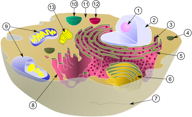
eukaryotic cell
A model of a eukaryotic cell (picture taken from On-Line Biology Book )
A eukaryotic cell has a nucleus, which is separated from the rest of the cell by a membrane. The nucleus contains chromosomes, which are the carrier of the genetic material (Section 3). There are internal membrane enclosed compartments within eukaryotic cells, called organelles, e.g., centrioles, lysosomes, golgi complexes, mitochondria among others (see picture above), which are specialised for particular biological processes. The mitochondria are found in all eukaryotes and are specialised for energy production (respiration). Chloroplasts are organelles found in plant cells which produce sugar using light. Light is the ultimate source of energy for almost all life on Earth. The area of the cell outside the nucleus and the organelles is called the cytoplasm. Membranes are complex structures and they are an effective barrier to the environment, and regulate the flow of food, energy and information in and out of the cell. There is a theory that mitochondria are prokaryotes living within eukaryotic cells.
An essential feature of most (prokaryote and eukaryote) living cells is their ability to grow in an appropriate environment and to undergo cell division. The growth of a single cell and its subsequent division is called the cell cycle. However, not all cells continually grow and divide, for example neurons only undergo an initial growth phase. Prokaryotes, particularly bacteria, are extremely successful at multiplying - it is likely that natural selection has favoured single celled organisms able to grow and divide quickly. Multicellular organisms typically begin life as a single cell, usually as a result of fusion of a male and a female sex cell (gametes). The single cell has to grow, divide and differentiate into different cell types to produce tissues and in higher eukarotyes, organs. Cell division and differentiation need to be controlled. Cancerous cells grow without control and can go on to form tumours. Development of single cells into complex organisms is in itself an area of study called developmental biology. This year’s  Nobel prize for Physiology or Medicine has been awarded to scientists for the discoveries of key regulators of the cell cycle.

Cells consist of molecules.

Human ecology and biology

Human ecologists
Human ecologists
Like nature, the human body must maintain a fragile balance in order to thrive. The smallest factor can effect human ecology. Human ecology can be thought of in two ways. There is human ecology in the sense that humans effect their environment and vice verse. However, their is also a fragile ecology inside the human body. This type of human ecology can be thought of as biology ecology. While humans can effect their natural environment the same cant always be said for our effect on our biology ecology. In some sense humans have control over their biology ecology. However, our environment and heredity can also directly effect the fragile balance in our bodies.
Getting lung cancer is an example that could be explained in either way. Human ecology might say that a person got lung cancer because they were a heavy smoker throughout their lifetime or where exposed to known cancer causing material such as asbestos. These days, people are aware of the effects of smoking on the human body and should act accordingly. By smoking or exposing ourselves to known carcinogens, we as the subject, caused ourselves to get lung cancer. However, lung cancer can also be explained through biology ecology. Certainly if the subject smoked and introduced a foreign substance into the human body, they caused the effect on the biology ecology of that subject. However, it could also be said that that persons biology effected the outcome of cancer. There are people that never smoke or purposefully exposed themselves to known carcinogens and they still get lung cancer. Those people may have not had a balance in their biology ecology and could complete no outside action (human ecology) to directly effect the outcome of lung cancer. We cannot always be held responsible for our biology or how it effects us. Regardless of the type of ecology, balance is always a necessity for an organism to thrive.
Human ecology may study the social reason for our smoking habit. It may also study the reason that society has allowed known carcinogens such as lead, to remain in our environment. In studying the social interaction between humans and their environment, human ecologists are able to find solutions to problems that are caused as a direct result of interaction between humans and their environment. Biology ecology may study biological factors in the human body that are out of our control. By studying the relationship between humans and their bodies, ecologists can sometimes find links in other types of ecology and find ways to produce a positive outcome.
Human health depends on many ecological factors. In many cases, humans have the ability to effect those factors. Humans can usually change their environmental factors through geography or through direct environmental contact. While we can not always change our biology, we can change environmental factors that will effect our biology. Humans have the ability to change many factors that effect us both directly and indirectly.